An innovative endoscopic operation to crush and remove deposits inside the liver was carried out at the Warsaw Medical University. It used the most modern equipment.
The first procedure of this type was performed in Poland by Dr. Jan Bertkevich and Dr. Swaomer Kosic of the Laboratory of General Endoscopy, Transplant and Hepatobiliary Surgery at Warsaw Medical University. The head of the clinic is Prof. Doctor Hub. med. Krzysztof Zieniewicz.
A patient who was reconstructed years ago after suffering damage during a cholecystectomy (cholecystectomy) was performed. In the postoperative period, there was a narrowing of the anastomosis of the intestinal tract and massive stones appeared inside the liver.
According to the PAP, the press officer of the Warsaw Medical University, Marta Wojtach, has failed attempts to perform an endoscopic cholangiography and cholangiography (ERCP), a combination of endoscopy and X-rays using a single-balloon bowel scope, has failed. Then it was decided to perform percutaneous biliary drainage (PTCD) with balloon anastomosis widening and external-internal drainage.
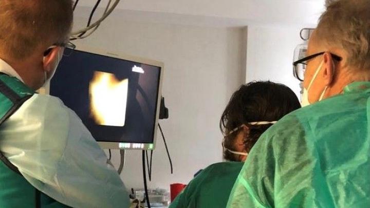
After a few weeks, the drainage duct was enlarged and the SpyGlass Discover biliary tract endoscopy was introduced with this method. Massive urolithiasis was confirmed in both liver lobes. The sediment was further divided into smaller pieces using an electro-hydraulic lithotripsy generator and probe. Then, using a basket and balloon, the sediment and its fragments were evacuated to the intestinal ring through a dilated anastomosis. “The patient is in very good general condition, free of diseases, returned home” – say the specialists of the Medical University of Warsaw in the information provided to the PAP.
SpyGlass Discover is a short, disposable endoscope used to explore the bile ducts during endoscopic or dermatological procedures. It allows you to bend the ends 180 degrees, so that you can reach all branches of the biliary tree with a diameter of at least 3 mm. Thanks to high-resolution imaging, even small changes in the mucous membrane of the bile duct and small deposits can be seen.
The working channel with a diameter of 1.2 mm allows the insertion of biopsy forceps, an electro-hydraulic lithotripsy (EHL) probe or laser lithotripsy, a tissue collection ring or a basket to evacuate deposits in the bile ducts. The procedure is performed under the control of sight, and not just an x-ray image, which significantly increases its effectiveness and safety.
The laboratory and clinic team intends to purchase such equipment for this procedure, which will allow other patients of the Warsaw Medical University to have constant access to the described technique. (PAP)
zbw / agt /

“Music specialist. Pop culture trailblazer. Problem solver. Internet advocate.”


